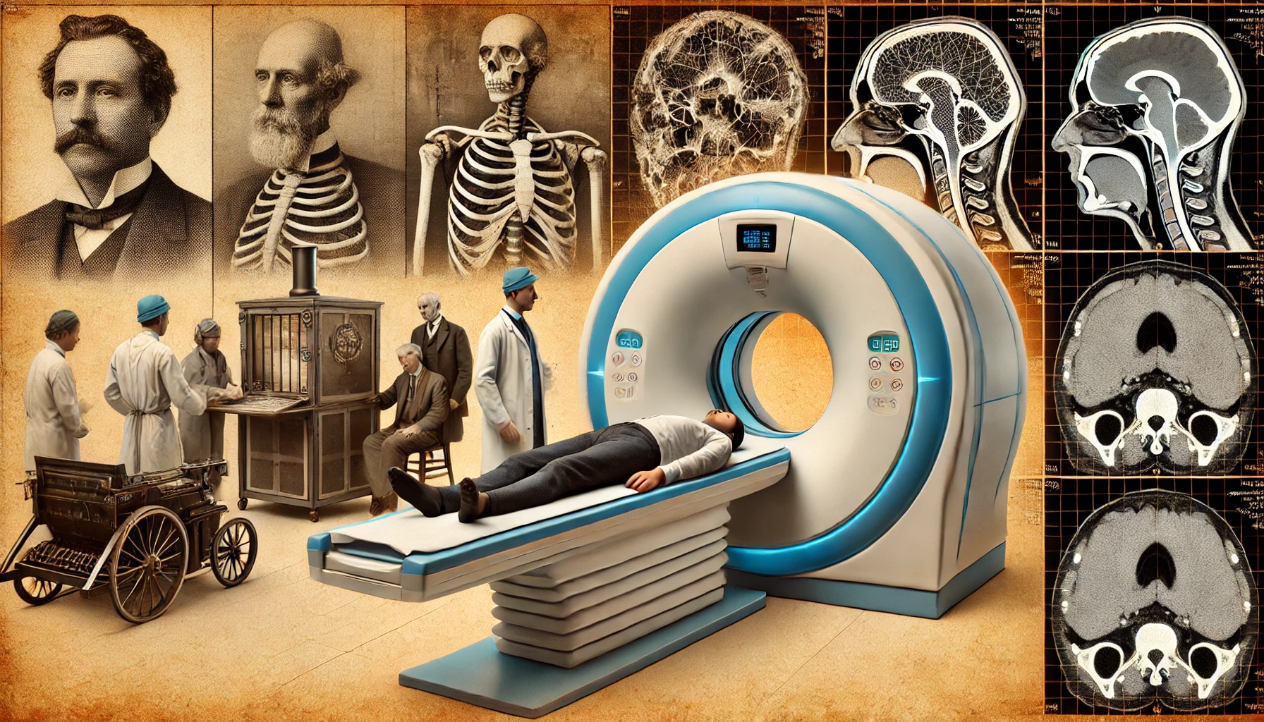
The discovery of X-rays in 1895 revolutionized diagnostic medicine, and the subsequent development of CT technology has played an important role in non-invasive and accurate diagnosis of the human body. CT is used in a wide variety of fields, and it requires both technological advances and ethical considerations.
The discovery of X-rays in 1895 revolutionized diagnostic medicine. Since then, X-ray photography technology has evolved into CT (computed tomography), which allows for the construction of three-dimensional images by taking cross-sectional images, allowing for accurate diagnosis of the inside of the body without dissection. These advances in diagnostic technology have had a profound impact not only in medicine, but also in many other fields. For example, archaeology has been able to determine the internal structure of artifacts without damaging them, and industry has been able to perform more precise quality inspections of products.
X-ray photographs are obtained by irradiating the human body with X-rays and sensitizing the transmitted X-rays onto film. Some of the irradiated X-rays are absorbed and scattered by the tissue, while the rest passes through the tissue and comes out the other side. The transmittance, which indicates the degree to which X-rays are transmitted, is the highest for air and decreases in the order of fat, water, and bone. The intensity of the transmitted X-rays also decreases with the lower the transmittance of the tissue through which they pass and the thicker the tissue, the weaker the X-rays. The intensity of these X-rays determines the degree of sensitization of the X-ray film, which results in a black-and-white image of the tissue. However, it is difficult to distinguish between tissues with similar transmittance, so X-ray photography is mainly used to examine bones or abnormal tissues that have a large difference in transmittance from other tissues. CT overcomes this limitation of X-rays.
CT reconstructs an image of a cross-section of the human body from the distribution of X-rays transmitted through the body. The CT machine has an X-ray generator on one side and several X-ray detectors on the other side. In the center of the CT machine, a bed with a person lying down enters, and X-rays from the X-ray generator penetrate the human body and are detected by the opposite X-ray detector. In this process, the X-ray transmittance from various angles is measured to obtain comprehensive image data. This helps to analyze the three-dimensional structure of the human body beyond a simple two-dimensional image.
The X-ray detector detects the intensity of the X-rays that have penetrated the human body, subtracting the amount that is attenuated by the air, to get the total amount of attenuated X-rays that have passed through the human tissue. This can be done by calculating the difference between the intensity of the X-rays through the air and the intensity of the X-rays through the tissue, which is known as the conversion factor, which is the total amount of attenuation due to scattering or absorption as the X-rays pass through the tissue in a particular direction. To obtain this value in multiple directions, the CT machine is rotated to obtain the equivalent value in each direction for the same cross-section, which is then used by the computer to reconstruct the cross-sectional image.
In CT, the back projection method is used to reconstruct the image. Back projection is a method of retracing the path traveled by X-rays in a certain direction and distributing the conversion values evenly along the path. The results of the back projection are added together by back-projecting the values obtained by rotating the CT machine in different directions, and the result is the result of the back projection. Back-projection results in the addition of values from multiple directions in tissues that are highly attenuated, such as bone, resulting in larger values than in other tissues.
By synthesizing the back-projected results, the distribution of tissues based on differences in transmission can be reconstructed in the image. By repeating the acquisition of different sections of the body as the CT machine moves in small increments, a series of cross-sectional images can be obtained, which can then be combined to create a three-dimensional image if necessary. This allows doctors to make more accurate diagnoses and treatment plans, which has a positive impact on patient outcomes.
Advances in CT technology have greatly improved the accuracy and efficiency of diagnosis. While early CT machines had limited resolution and acquisition time, modern CT machines are able to acquire high-resolution images in a fraction of the time. This allows for rapid diagnosis, even in emergency situations, which can be crucial in saving a patient’s life. For example, in the case of stroke patients, CT can quickly identify brain bleeding and initiate appropriate treatment.
CT technology also plays an important role in cancer diagnosis and treatment. Because cancer cells have a different density than normal tissue, CT can be used to detect them early, and it can also be used to monitor the progress of treatment. This contributes to increasing the survival rate of patients and maximizing the effectiveness of treatment.
The applications of CT technology extend beyond medicine to various fields. For example, in veterinary medicine, CT can be used to accurately identify and diagnose the internal structures of animals. In forensic medicine, CT is also used to analyze the internal structures of corpses to determine the cause of death. As you can see, CT technology plays an important role in various fields, and its applications are expected to expand in the future.
At the same time, it is important to consider the ethical implications of advances in CT technology. Since CT scans use X-rays, radiation dose can be an issue. Therefore, it is important to perform CT scans only when necessary and with minimal exposure. In addition, privacy and data management using CT data are also important issues. Continued research and regulation are needed to address these issues.
In conclusion, the development of X-ray and CT technology has revolutionized medical diagnosis and treatment and plays an important role in various fields. Their applications will continue to expand with the advancement of technology, and they will contribute to improving the quality of human life.
 I’m a blog writer. I want to write articles that touch people’s hearts. I love Coca-Cola, coffee, reading and traveling. I hope you find happiness through my writing.